
Dîroka serîlêdanê û rola mîkroskopên cerrahî di neuroşîrurjiyê de
Di dîroka neştergeriya mejî de, sepandinamîkroskopên cerrahîsemboleke şoreşger e, ku ji serdema kevneşopî ya neştergeriya neuro ya pêkanîna emeliyatê di bin çavê tazî de ber bi serdema nûjen a neştergeriya neuro ya pêkanîna emeliyatê di bin...mîkroskûpKê û kengî kirmîkroskopên xebitandinêdest bi karanîna di cerrahîya mejî de bikin? Rola wê çi yemîkroskopa cerrahîdi pêşxistina neştergeriya mejî de lîstiye? Bi pêşketina zanist û teknolojiyê re, wêMîkroskopa xebitandinêbi hin alavên pêşketîtir were guhertin? Ev pirsek e ku divê her cerrahê mejî jê haydar be û teknolojiya herî dawî û amûrên xwe di warê cerrahîya mejî de bi kar bîne, da ku baştirkirina jêhatîyên cerrahîya mejî pêş bixe.
1. Dîroka Serlêdanên Mîkroskopê di Qada Tibbî de
Di fîzîkê de, lensên çavikên çavan lensên konveks in ku xwedî avahiyek yekane ne ku bandorek mezinkirinê hene, û mezinbûna wan sînordar e, ku wekî lensên mezinkirinê têne zanîn. Di sala 1590 de, du kesên Holandî du plakayên lensên konveks di hundurê bermîleke silindirî ya zirav de saz kirin, bi vî rengî yekem amûra mezinkirinê ya avahiya kompozît a cîhanê îcad kirin:mîkroskûpPaşê, avahiya mîkroskopê bi berdewamî baştir bû, û mezinbûn bi berdewamî zêde bû. Di wê demê de, zanyar bi piranî vê yekê bi kar dianîn.mîkroskopa pêkhatîji bo çavdêriya avahiyên piçûk ên ajalan û nebatan, wek avahiya şaneyan. Ji nîvê heta dawiya sedsala 19an, camên mezinkirinê û mîkroskop hêdî hêdî di warê bijîşkî de hatine sepandin. Di destpêkê de, cerrahan ji bo emeliyatê camên mezinkirinê yên bi şêweya camên çavan bi avahiyek yekane bikar anîn ku dikarin li ser pira poz werin danîn. Di sala 1876an de, bijîşkê Alman Saemisch yekem emeliyata "mîkroskopîk" a cîhanê bi karanîna camek mezinkirinê ya camên çavan a tevlihev pêk anî (cureya emeliyatê nayê zanîn). Di sala 1893an de, pargîdaniya Alman Zeiss îcad kirmîkroskopa dûrbîn, bi giranî ji bo çavdêriya ceribandinî di laboratuarên bijîşkî de, û her weha ji bo çavdêriya birînên kornea û odeya pêşiyê di warê oftalmolojiyê de tê bikar anîn. Di sala 1921-an de, li ser bingeha lêkolînên laboratîfê yên li ser anatomiya guhê hundurîn ê ajalan, otolaringologê Swêdî Nylen guhekî sabît bikar anî.mîkroskopa cerrahî ya yekalîji hêla wî ve hatîye sêwirandin û çêkirin da ku emeliyata otîta navîn a kronîk li ser mirovan bike, ku mîkrocerrahiyek rastîn bû. Salek şûnda, bijîşkê payebilind ê Nylen, Hlolmgren, destpêkek da nasandin.mîkroskopa cerrahî ya bînokulerji hêla Zeiss ve di odeya emeliyatê de hatî çêkirin.
ZûtirînMîkroskopên xebitandinêgelek kêmasî hebûn, wek mînak aramiya mekanîkî ya nebaş, nekarîna tevgerê, ronîkirina tewereyên cuda û germbûna lenza objektîfê, qada mezinkirina neştergeriyê ya teng, û hwd. Ev hemû sedem in ku sepandina berfirehtir amîkroskopên cerrahîDi sî salên pêş de, ji ber têkiliya erênî di navbera cerrahan ûhilberînerên mîkroskopê, performansamîkroskopên cerrahîbi berdewamî baştir dibû, ûmîkroskopên cerrahî yên bînokuler, mîkroskopên li ser banî hatine danîn, lensên zoomê, ronîkirina çavkaniya ronahiyê ya koaksiyal, destên artikulkirî yên elektronîkî an jî bi zexta avê kontrolkirî, kontrola pedalê lingan, û hwd. li pey hev hatin pêşxistin. Di sala 1953an de, şîrketa Almanî Zeiss rêze lensên pispor hilberand.mîkroskopên cerrahî ji bo otolojiyê, bi taybetî ji bo emeliyatên li ser birînên kûr ên wekî guhê navîn û hestiyê demkî minasib e. Dema ku performansamîkroskopên cerrahîberdewam baştir dibe, zîhniyeta cerrahan jî bi berdewamî diguhere. Bo nimûne, bijîşkên Alman Zollner û Wullstein destnîşan kirin kumîkroskopên cerrahîdivê ji bo emeliyata şekildana parzûna tympanîk were bikar anîn. Ji salên 1950-an vir ve, oftalmologan hêdî hêdî pratîka karanîna tenê mîkroskopan ji bo muayeneyên oftalmîk guhertin û destnîşan kirin.mîkroskopên otocerrahînav cerrahîya çavan. Ji wê demê ve,Mîkroskopa xebitandinêdi warên otolojî û oftalmolojiyê de bi berfirehî hatiye bikar anîn.
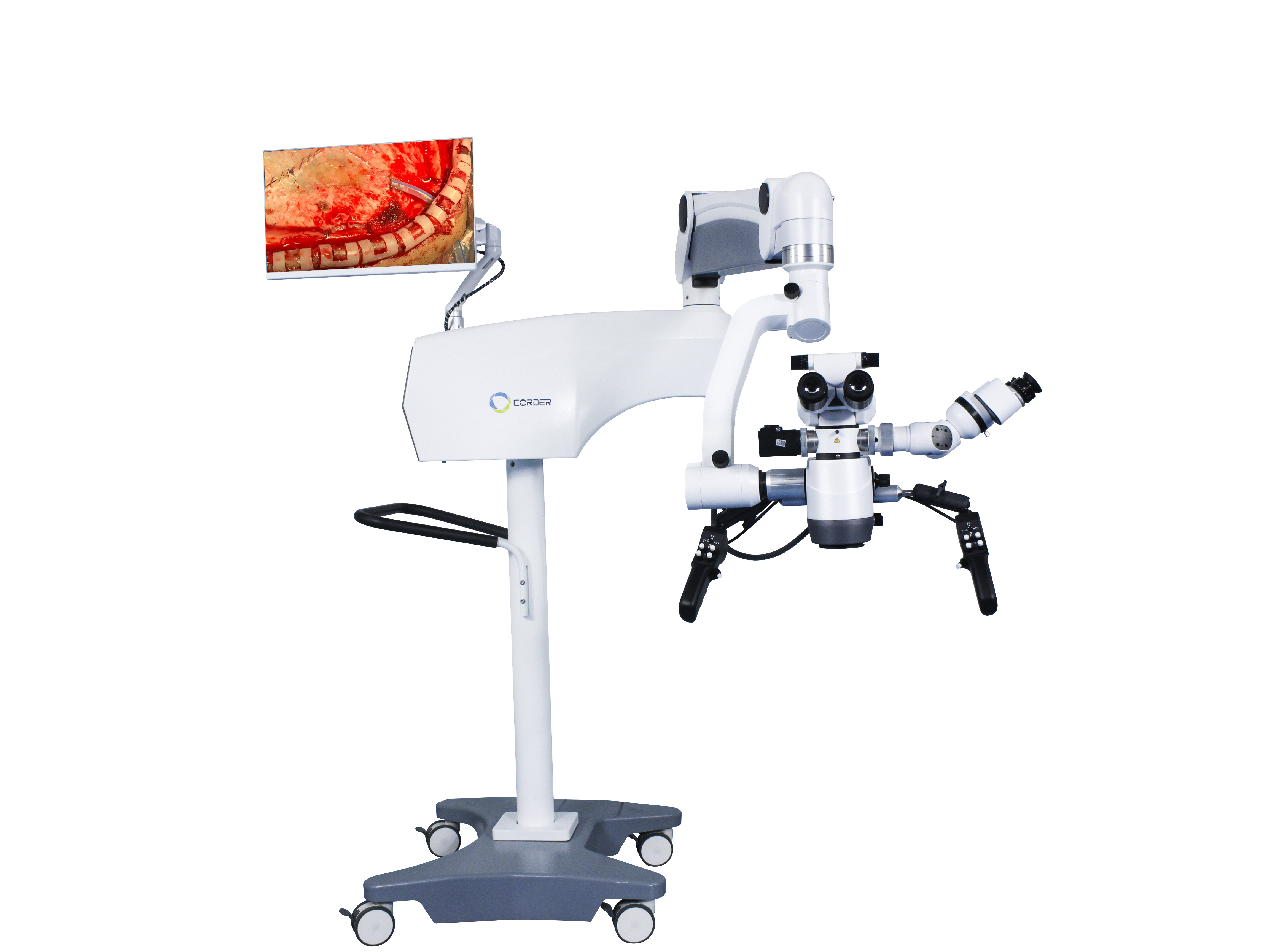
2、Sepandina mîkroskopa cerrahî di neuroşîrurjiyê de
Ji ber taybetmendiya neştergeriya mejî, sepandinamîkroskopên cerrahî di neuroşîrurjiyê dehinekî derengtir e ji otolojî û oftalmolojiyê, û cerrahên mejî bi awayekî çalak vê teknolojiya nû fêr dibin. Di wê demê de,bikaranîna mîkroskopên cerrahîbi piranî li Ewropayê bû. Oftalmologê Amerîkî Perrit yekem car da nasînmîkroskopên cerrahîji Ewropayê bo Dewletên Yekbûyî yên Amerîkayê di sala 1946an de, bingehek ji bo bikaranîna neurocerrahên Amerîkî danî.Mîkroskopên xebitandinê.
Ji perspektîfa rêzgirtina nirxa jiyana mirovan, divê her teknolojiyek, alav an amûrên nû yên ku ji bo laşê mirovan têne bikar anîn, ceribandinên pêşîn ên li ser heywanan û perwerdehiya teknîkî ya operatoran derbas bikin. Di sala 1955an de, cerrahê mejî yê Amerîkî Malis bi karanîna...mîkroskopa cerrahî ya bînokulerKurze, cerrahê mejî li Zanîngeha Başûrê Kalîforniyayê li Dewletên Yekbûyî, piştî ku emeliyata guh di bin mîkroskopê de temaşe kir, salekê teknîkên emeliyatê yên karanîna mîkroskopê di laboratuwarê de fêr bû. Di Tebaxa 1957an de, wî bi serkeftî emeliyateke newroma akustîk li ser zarokekî 5 salî bi karanîna...mîkroskopa emeliyata guh, ku yekem emeliyata mîkrocerrahî ya cîhanê bû. Di demek kurt de piştî wê, Kurze bi serkeftî anastomoza nerva rû ya binzimanî li ser zarok pêk anî bi karanînamîkroskopa cerrahî, û başbûna zarok pir baş bû. Ev duyemîn emeliyata mîkrocerrahî li cîhanê bû. Piştre, Kurze kamyonan bikar anî da kuMîkroskopên xebitandinêji bo mîkrocerrahîya neurocerrahîyê bo cîhên cûrbecûr hatine şandin, û bi tundî karanînamîkroskopên cerrahîbo cerrahên din ên mejî. Piştre, Kurze emeliyata qipkirina anevrîzma mejî bi karanînamîkroskopa cerrahî(mixabin, wî tu gotar neweşand). Bi piştgiriya nexweşek newraljiya sêgoşeyî ku wî derman dikir, wî di sala 1961an de yekem laboratuwara mîkrocerrahiyê ya bingeha serî ya cîhanê ava kir. Divê em her gav beşdariya Kurze di mîkrocerrahiyê de bi bîr bînin û ji wêrekiya wî ya qebûlkirina teknolojiyên nû û ramanên nû fêr bibin. Lêbelê, heta destpêka salên 1990î, hin cerrahên neuro li Çînê qebûl nedikirin.Mîkroskopên neurocerrahîji bo emeliyatê. Ev ne pirsgirêkek bû biMîkroskopa neurocerrahîbi xwe, lê pirsgirêkek bi têgihîştina îdeolojîk a cerrahên mejî re ye.
Di sala 1958an de, cerrahê mejî yê Amerîkî Donaghy li Burlington, Vermont, yekem laboratuwara lêkolîn û perwerdehiya mîkrocerrahiyê ya cîhanê ava kir. Di qonaxên destpêkê de, ew ji serokên xwe re rastî tevlihevî û zehmetiyên darayî jî hat. Li akademiyê, wî her gav xeyal dikir ku damarên xwînê yên kortikalê veke da ku rasterast tromb ji nexweşên bi tromboza mejî derxe. Ji ber vê yekê wî bi cerrahê damaran Jacobson re li ser lêkolînên heywanan û klînîkî hevkariyê kir. Di wê demê de, di bin şert û mercên çavê tazî de, tenê damarên xwînê yên piçûk ên bi qûtra 7-8 mîlîmetre an jî zêdetir dikarin werin dirûtin. Ji bo ku anastomoza serî-bi-serî ya damarên xwînê yên ziravtir pêk were, Jacobson pêşî hewl da ku camek mezinkirinê ya bi şêwaza çavik bikar bîne. Di demek kurt de, wî bi bîr xist ku karanînamîkroskopa cerrahî ya otolaryngolojiyêji bo neştergeriyê dema ku ew bijîşkê niştecî bû. Ji ber vê yekê, bi alîkariya Zeiss li Almanya, Jacobson mîkroskopa neştergeriyê ya du operatorî sêwirand (Dîploskop) ji bo anastomoza damarî, ku dihêle du cerrah di heman demê de emeliyatê bikin. Piştî ceribandinên berfireh ên li ser heywanan, Jacobson gotarek li ser anastomoza mîkrocerrahî ya kûçikan û damarên ne-karotîd (1960) weşand, bi rêjeya vebûna 100% ya anastomoza damarî. Ev gotarek bijîşkî ya şoreşger e ku bi neurcerahiya mîkrocerrahî û cerrahiya damarî ve girêdayî ye. Jacobson her weha gelek amûrên mîkrocerrahî, wekî mîkro maqes, girtina derziyên mîkro, û destên mîkro amûran sêwirand. Di sala 1960 de, Donaghy bi serkeftî trombektomiyek birîna damara mejî di binmîkroskopa cerrahîji bo nexweşek bi tromboza mejî. Rhoton ji Dewletên Yekbûyî di sala 1967an de dest bi lêkolîna anatomiya mejî di bin mîkroskopê de kir, pêşengiya qadek nû ya anatomiya mîkrocerrahî kir û beşdarîyên girîng di pêşxistina mîkrocerrahîyê de kir. Ji ber avantajênmîkroskopên cerrahîû bi pêşxistina amûrên mîkrocerrahî, bêtir û bêtir cerrah hez dikin ku wan bikar bîninmîkroskopên cerrahîji bo neştergeriyê. Û gelek gotarên têkildar li ser prosedurên mîkrocerrahî weşandin.
3, Bikaranîna mîkroskopa cerrahî di neuroşîrurjiyê de li Çînê
Wekî welatparêzekî Çînî yê derveyî welêt li Japonyayê, Profesor Du Ziwei yekem çînîya navxweyî bexş kir.mîkroskopa neurocerrahîû têkildaramûrên mîkrocerrahîdi sala 1972an de ji bo Beşa Neurşîrurjiya Mejî ya Nexweşxaneya Girêdayî Koleja Bijîşkî ya Suzhou (niha Beşa Neurşîrurjiya Mejî ya Nexweşxaneya Yekem a Girêdayî Zanîngeha Suzhou) hate şandin. Piştî vegera xwe ya Çînê, wî pêşî emeliyatên mîkrocerrahî yên wekî anevrîzmayên mejî yên hundirîn û menengîoma pêk anîn. Piştî ku li ser hebûnamîkroskopên neurocerrahîû amûrên mîkrocerrahî, Profesor Zhao Yadu ji Beşa Neurşîrurjiya Mejî ya Nexweşxaneya Yiwu ya Pekîn serdana Profesor Du Ziwei ji Koleja Bijîşkî ya Suzhou kir da ku karanînamîkroskopên cerrahîProfesor Shi Yuquan ji Nexweşxaneya Şanghayê Huashan bi xwe serdana beşa Profesor Du Ziwei kir da ku prosedurên mîkrocerrahîyê bibîne. Di encamê de, pêlek ji danasîn, fêrbûn û sepandinaMîkroskopên neurocerrahîli navendên sereke yên neştergeriya mejî li Çînê derket holê, ku destpêka mîkroneştergeriya mejî ya Çînê nîşan da.
4、Bandora Emeliyata Mîkrocerrahîyê
Ji ber bikaranînamîkroskopên neurocerrahî, emeliyatên ku bi çavê tazî nayên kirin di şert û mercên mezinkirina 6-10 caran de gengaz dibin. Bo nimûne, kirina emeliyata tumora hîpofîzê bi rêya sînusa etmoidal dikare bi ewlehî tumorên hîpofîzê tespît bike û rake di heman demê de rijêna hîpofîzê ya normal diparêze; Emeliyatên ku bi çavê tazî nayên kirin dikarin bibin emeliyatên çêtir, wekî tumorên stûna mêjî û tumorên navmedullary yên stûna piştê. Akademîsyen Wang Zhongcheng berî ku ameliyata anevrîzmaya mêjî bikar bîne, rêjeya mirinê ji bo emeliyata anevrîzmaya mêjî %10.7 bû.mîkroskopa neurocerrahîPiştî bikaranîna mîkroskopê di sala 1978an de, rêjeya mirinê daket 3.2%. Rêjeya mirinê ya emeliyata malformasyona arteriovenoz a mejî bêyî bikaranîna mîkroskopê.mîkroskopa cerrahî%6.2 bû, û piştî sala 1984an, bi karanînamîkroskopên neurocerrahî, rêjeya mirinê daket 1.6%. Bikaranînamîkroskopa neurocerrahîdihêle ku tumorên hîpofîzê bi rêbazek transnazal a transsphenoidal a kêm-dagirker bêyî hewcedariya kraniotomiyê werin dermankirin, rêjeya mirina neştergerî ji %4.7 kêm dike bo %0.9. Bi neştergeriya çavê giştî ya kevneşopî bidestxistina van encaman ne mumkin e, ji ber vê yekêmîkroskopên cerrahîsembola cerrahîya mejî ya nûjen in û bûne yek ji alavên cerrahî yên neçar û neguhêrbar di cerrahîya mejî ya nûjen de.
Dema şandinê: Kanûn-09-2024


